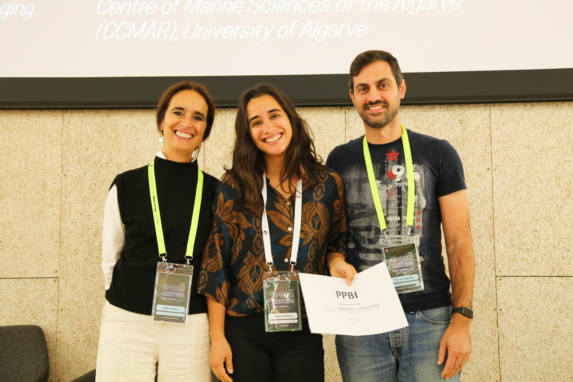
Our microscopy platform has just won first prize in the microscopy image competition organised by PPBI – Portuguese Platform of BioImaging, in partnership with the conference SPAOM (Spanish & Portuguese Advanced Optical Microscopy Meeting).
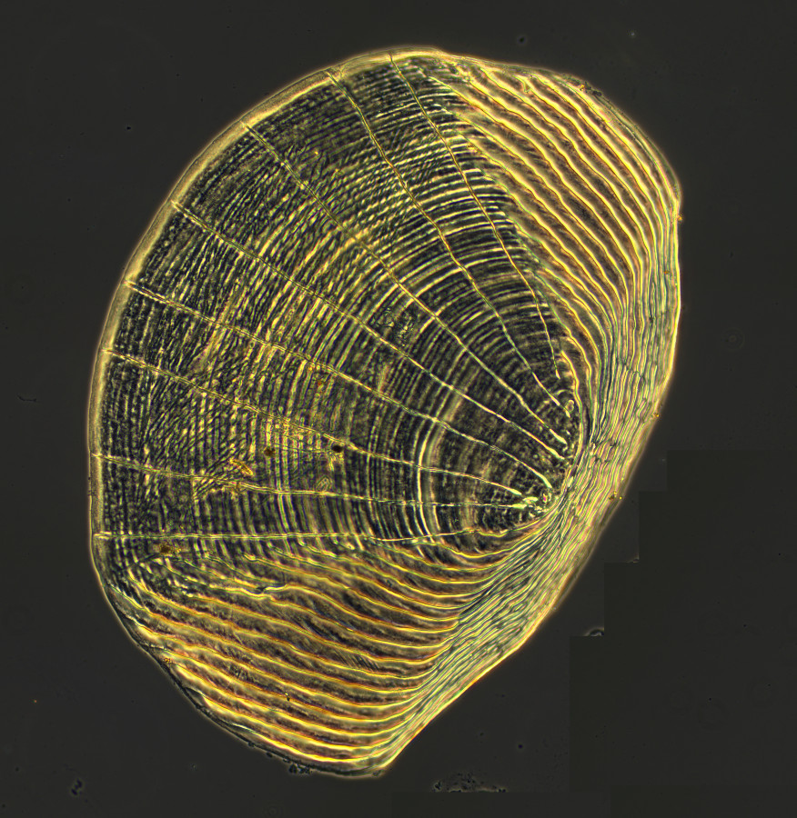
The winning image, captured by Carina Mónico, head of the microscopy platform, reveals the striking scientific beauty of a zebrafish scale (Danio rerio) — a model organism widely used in biomedical research, developmental biology, and ecology.

“What at first glance appears to be a rhythmic engraving or a delicate scallop shell is actually a zebrafish scale. The sample was prepared without additional staining in order to preserve its natural structure, observed with a 10x lens and colour camera. The image highlights the concentric lines typical of scales, which record the fish's growth cycles in a similar way to tree rings.” — Carina Mónico, CCMAR Imaging Platform
The sample used was kindly provided by Gil Martins, our researcher integrated in the Bioskel(Comparative, Adaptive and Functional Skeletal Biology) group, which reveals the spirit of collaboration that characterises the research at our centre.
The image was acquired with the DM6 B microscope (Leica) in dark field, a technique that enhances contrast and allows structural details to be revealed in an artistic and scientific way.
The PPBI Image Contest celebrates art and creativity in science, challenging researchers from Portuguese institutions to showcase the aesthetic side of their microscopic discoveries. The winning photograph highlights not only the technical excellence of the CCMAR team, but also the potential of bioimaging to bring science closer to the public.